


High‑Performance Flexible Superhydrophobic Silver Nanowire Arrays for Ultra‑Sensitive Surface‑Enhanced Raman Scattering
Abstract
We present a flexible, superhydrophobic substrate composed of silver‑nanowire (AgNW) arrays decorated with silver nanoparticles (AgNPs) embedded in shape‑memory polyurethane (SMPU). The fabrication strategy—interfacial self‑assembly, electrochemical copper deposition, galvanic AgNP growth, and perfluorodecanethiol (PFDT) functionalization—creates a hierarchical micro‑/nanostructure that both traps analyte molecules and amplifies electromagnetic hotspots. Using Rhodamine B (RB) as a probe, the substrate achieves a detection limit of 10⁻¹⁰ M, a 1.5‑fold intensity gain over non‑hydrophobic counterparts, and excellent reproducibility (RSD < 24 %). These results demonstrate that combining flexibility, superhydrophobicity, and plasmonic enhancement yields a robust, sensitive SERS platform suitable for real‑world sensing applications.
Introduction
Surface‑enhanced Raman scattering (SERS) has emerged as a premier technique for trace and single‑molecule detection, owing to its extraordinary signal amplification through localized surface plasmon resonances (LSPR). Hotspots—regions of concentrated electromagnetic fields—typically appear at nanometer‑scale gaps, sharp tips, and high‑curvature points, enabling enhancements up to |E|⁴. While colloidal nanoparticles and roughened electrodes offer simplicity, they suffer from poor reproducibility. Top‑down nanofabrication delivers high quality but at prohibitive cost and complexity. Self‑assembly, in contrast, provides a scalable, cost‑effective route to well‑ordered plasmonic architectures with controllable interparticle spacing, avoiding lithographic drawbacks.
Superhydrophobicity further augments SERS by confining analytes to sub‑micron areas through capillary pinning, thereby concentrating target molecules and boosting detection limits. Prior efforts have employed lotus‑like surfaces or polymer coatings to impart low‑surface‑energy, but achieving stable, high‑roughness nanostructures remains challenging. Flexible substrates expand applicability by enabling conformal contact with irregular surfaces and facilitating sample collection. Shape‑memory polyurethane (SMPU) offers shape recovery and tunable mechanical compliance, making it an attractive matrix for integrating plasmonic nanostructures.
In this work, we develop a multifunctional SERS substrate that synergistically combines aligned AgNW arrays, AgNP decoration, SMPU flexibility, and PFDT‑induced superhydrophobicity. The resulting platform delivers ultralow detection limits, excellent uniformity, and long‑term stability.
Methods
Materials
Perfluorodecanethiol (PFDT) (Sigma‑Aldrich), AgNO₃ and CuSO₄ (analytical grade, Beijing Chemical Reagents), AgNW aqueous suspension (diameter 300 nm, length 30 µm, Haoxi Research Nanomaterials), and SMPU synthesized according to literature [31].
Aligned AgNW Film Fabrication
AgNWs were dispersed (5 mg mL⁻¹) and dropped onto a chloroform/water interface. Dropwise acetone addition induced self‑assembly, yielding a mirror‑like monolayer. The film was transferred to precleaned silicon chips and hot‑pressed onto SMPU (≈ 50 µm thickness) to form composite S0.
Copper‑Decorated AgNW Films
The S0 film was immersed in a mixed electrolyte (70 g L⁻¹ CuSO₄, 200 g L⁻¹ H₂SO₄, 50 ppm HCl, 1 ppm Bis‑(3‑sodiumsulfopropyl disulfide), PEG 6000, 1 ppm Janus Green) and electroplated at 0.1 A for 5–60 s. Samples S1–S4 correspond to deposition times of 5, 15, 30, and 60 s.
AgNP Decoration
Cu‑decorated films were immersed in 1 × 10⁻³ M AgNO₃ for 1 min to induce galvanic replacement, forming AgNPs on AgNWs. The resulting AgNWs@AgNPs films were rinsed and N₂‑dried.
Superhydrophobic Functionalization
Films were immersed in 5 mM PFDT in 1:1 ethanol/hexane for 15 h, then washed with ethanol and dried. This step imparts low surface energy and a hierarchical roughness necessary for superhydrophobicity.
Characterization
SEM (JEOL JSM‑7001F) examined morphology; UV–vis (Shimadzu UV‑2450) measured LSPR; XRD (Bruker X’Pert) confirmed crystal phases. Static water contact angles were measured with a goniometer (JC2000D1). SERS spectra were collected on a HORIBA LabRAM HR 800 (633 nm, 1.7 mW, 20 s, 1 µm spot). All experiments were performed in triplicate.
Results and Discussion
Fabrication Overview
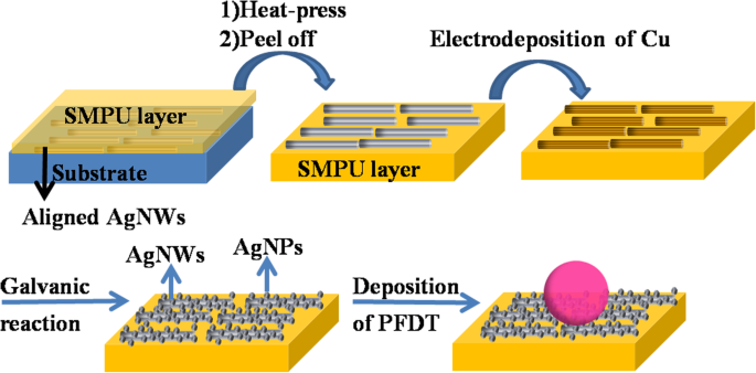
Figure 1 illustrates the three‑step process: (1) interfacial assembly of aligned AgNWs and integration into SMPU; (2) copper electroplating followed by galvanic AgNP growth; (3) PFDT surface modification.
Morphology and Structural Analysis
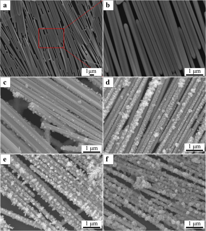
SEM images (Fig. 2a,b) reveal a highly ordered AgNW monolayer with minimal defects. Subsequent Cu deposition introduces surface roughness, and AgNPs form uniformly across the nanowire network (Fig. 2c–f). Particle size increases with deposition time, allowing precise control over hotspot density.
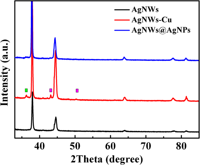
XRD patterns (Fig. 3) confirm the presence of face‑centered cubic Ag and the disappearance of Cu peaks after galvanic replacement, indicating complete transformation to AgNPs without residual Cu contamination.
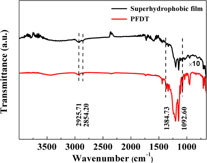
FT‑IR spectra (Fig. 4) demonstrate successful PFDT grafting, evidenced by red‑shifted CF vibrations and the absence of free PFDT peaks, confirming an ordered monolayer.
Superhydrophobicity Assessment
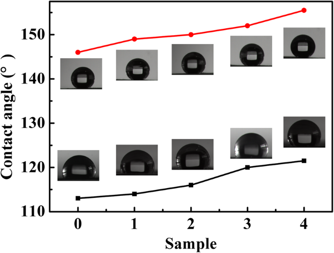
Static contact angles ranged from 113°–121° for AgNWs and AgNWs@AgNPs, increasing to 155° after PFDT functionalization (Fig. 5). The enhanced roughness and low surface energy produce a robust superhydrophobic state that pinches droplets into micrometer‑scale spots.
Concentrating Effect
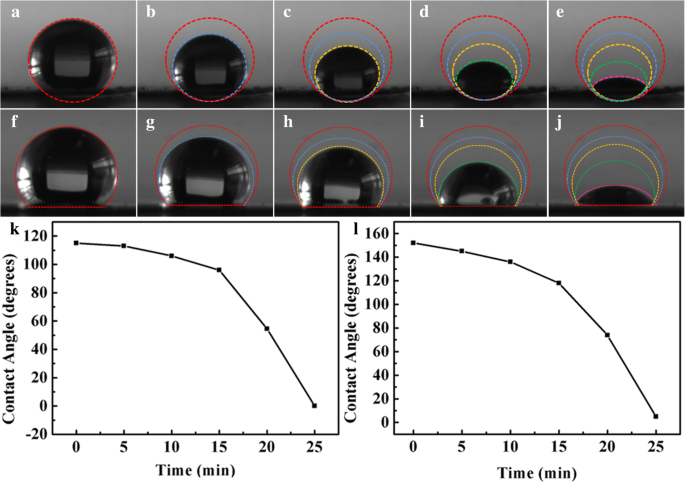
Evaporation of a 5 µL RB droplet on the superhydrophobic surface produced a confined spot (~0.60 mm²), five times smaller than the ~3.2 mm² spot on the hydrophilic AgNWs@AgNPs film (Fig. 6). This dramatic reduction in spot area translates into a higher local analyte concentration, directly benefiting SERS sensitivity.
Optical Properties
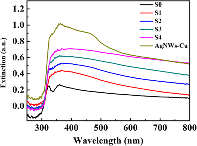
UV‑vis spectra (Fig. 7) show distinct LSPR peaks at 323 nm and 352 nm for AgNWs, broadening and shifting to 450 nm after AgNP growth, confirming successful plasmonic coupling.
SERS Performance
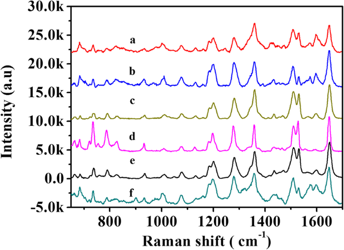
Using 10⁻⁵ M RB, the AgNWs@AgNPs film (S4) produced a marked Raman enhancement compared to the pristine film (S0). The superhydrophobic counterpart (S4‑SH) further amplified the signal by ~1.5×, attributable to analyte concentration and sustained hotspot accessibility (Fig. 8a,b).
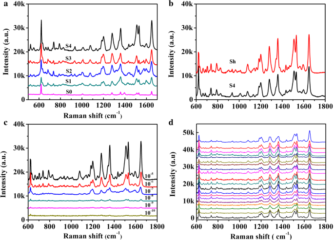
Detection limits reached 10⁻¹⁰ M RB, with characteristic peaks remaining discernible down to this concentration (Fig. 8c). The relative standard deviation across 20 random sites was < 24 %, evidencing excellent uniformity (Table 1).
Stability
SERS signals remained stable over 24 h of storage, with peak intensities varying by less than 5 %, indicating robust substrate performance (Fig. 9).
Conclusion
We have engineered a flexible, superhydrophobic SERS substrate by integrating aligned AgNW arrays with AgNP decoration, SMPU embedding, and PFDT functionalization. The hierarchical micro‑/nanostructure provides abundant hotspots, while superhydrophobicity concentrates analytes into micrometer‑scale regions, collectively enabling detection of Rhodamine B down to 10⁻¹⁰ M. The substrate exhibits excellent uniformity, reproducibility, and stability, positioning it as a promising platform for practical, on‑site chemical sensing.
Abbreviations
- AgNPs
- Silver nanoparticles
- AgNWs
- Silver nanowires
- AgNWs@AgNPs
- Silver nanoparticles decorated aligned silver nanowires
- CA
- Static contact angle
- PFDT
- Perfluorodecanethiol
- RB
- Rhodamine B
- SEM
- Scanning electron microscope
- SERS
- Surface‑enhanced Raman scattering
- SMPU
- Shape memory polyurethane
- XRD
- X‑ray diffraction
Nanomaterials
- pH‑Responsive Ag@Polyacryloyl Hydrazide Nanoparticles: A Smart, Ultra‑Sensitive SERS Substrate for Trace Analysis
- High‑Performance Flexible Transparent Electrodes from Silver Nanowires with Tailored Aspect Ratios
- Highly Sensitive SERS Substrates from Aligned, Chemically Etched Silver Nanowire Monolayers
- Ultra-Sensitive Large-Scale SERS Substrates: Silver Nanowire Thin Films via Microliter-Scale Solution Coating
- Epitaxial Growth of High‑Quality SrGe₂ Thin Films on Ge Substrates via Reactive Deposition Epitaxy
- Advances in Synthesis and Applications of Silver Nanostructures
- Graphene Oxide–Silver Nanoparticle Nanocomposites: A Potent Antibacterial and Antifungal Agent
- Large-Scale Dendritic Silver Nanostructures: Controlled Morphology for Enhanced Catalysis and SERS Performance
- Raman Spectroscopy: Foundations, Innovations, and Emerging Frontiers
- Scalable, Patternable Nano‑Dot Arrays on ITO via Electrolysis for High‑Performance SERS