


3D‑Printed Surgical Models Enable Historic Separation of Rare Head‑Fused Twins
3D printing is reshaping medicine, and its impact is most evident in the growing use of anatomically accurate surgical models. In a landmark Israeli operation, these models were pivotal in separating craniopagus twins—children conjoined at the skull—who had a very low chance of survival.
The Rarity of Craniopagus Twins
Craniopagus twins occur in roughly 1 in 2.5 million births worldwide, with only about 5 % of conjoined twins fused at the head. This rare condition involves complex cranial sinuses, vascular networks, and intricate neuroanatomy, making surgical planning exceptionally challenging.
At one year old, the twins were considered a long‑shot, yet they became the first successfully separated craniopagus twins in Israel’s history—and one of only a handful worldwide.
Critical Role of 3D‑Printed Models
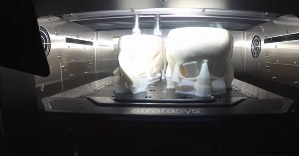
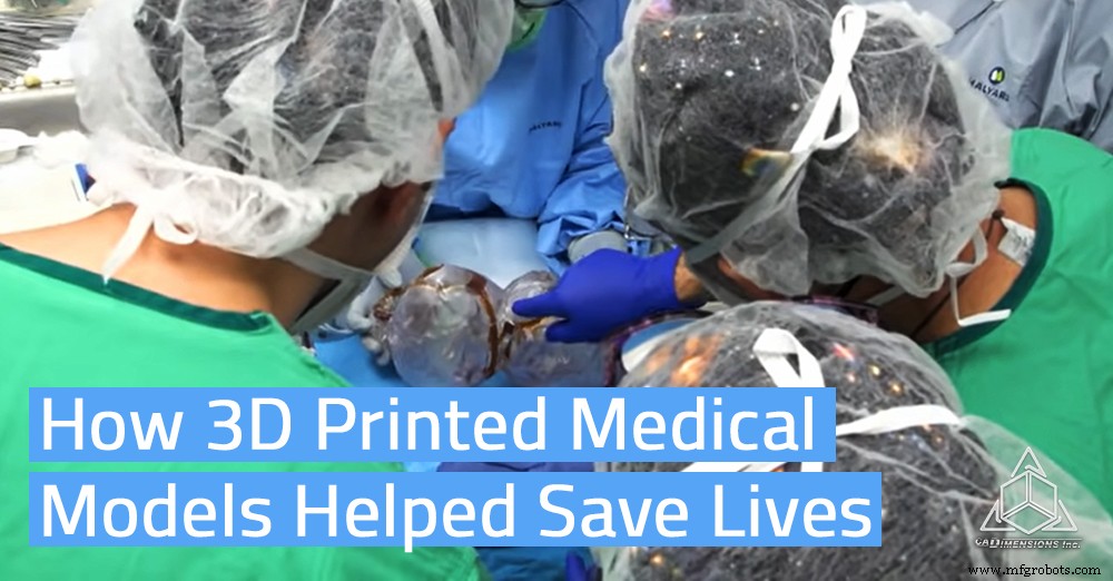
Leading the effort was Dr. Noor Ul wase Jeelani, a Kashmiri‑born pediatric neurosurgeon, who directed a multidisciplinary team at Soroka‑University in Israel. The pre‑operative plan integrated tools from 3D4OP, Surgical Theater, and Stratasys, while scans from CT and MRI machines were processed with Materialise 3‑Matic medical software to produce life‑like models.
These highly accurate replicas allowed surgeons to identify blood vessels, dura mater, skin, and bone in a tactile format identical to what they would encounter during surgery. The models could be positioned to match the twins’ orientation in the operating room, enabling rehearsal for both surgical and anesthesiology teams.
Successful 12‑Hour Surgery
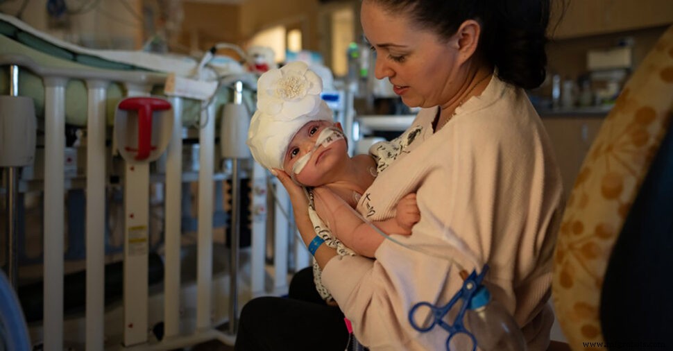
The procedure spanned 12 hours, after which Dr. Michael Gideon, director of pediatric neurosurgery at Soroka University Medical Centre, reported the twins were “conscious, alert, and neurologically intact.” While the postoperative period remains demanding, early indicators suggest both children can lead fully normal lives.
The Promise of 3D Printing in Surgery
3D printing enabled the medical team to anticipate how the human body would respond and to practice on a model that precisely reflected the twins’ unique anatomy. This case illustrates the transformative potential of additive manufacturing to improve surgical precision and patient outcomes across the globe.
Explore UC Davis Health’s 4‑Part Series
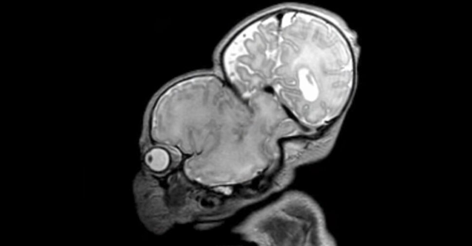
3D Models Are Printed From MRI & CT Scan Data
See Why Nothing Comes Close To The Repeatability & Consistency Between Samples Of 3D Printed Surgical Models
3D printing
- SPICE Diode Modeling: A Practical Guide to Accurate Simulation
- What to Make of 3D‑Printed Firearms: Ethics, Technology, and Public Policy
- 3D‑Printed Drone Enables Rapid, Cost‑Effective Data Collection in Antarctica
- Skateboard Part 1: Building Durable 3D‑Printed Wheels
- Revolutionizing 3D Printing: Precise Joinery for Effortless Assembly
- Introducing Inconel 625 for 3D Printing – A Game‑Changing Nickel‑Based Superalloy
- Markforged Introduces 3D‑Printed Copper – Unlocking High‑Conductivity Parts
- Digital Twins: Precision Data Models Are the Key to Successful Digital Transformation
- Are 3D Printed Parts Really Strong?
- Silencio: A 3D-Printed Tactile Poetry Book for Sighted and Blind Readers