


New 3D‑Printed Bio‑Ink Corneas Could Cut Donor Shortages – 10‑Minute Fabrication Achieved
- 3‑minute 3D‑printed corneas could dramatically reduce dependence on donor tissue.
- Printed entirely in under 10 minutes using a novel bio‑ink.
- Clinical translation may still take several years, as extensive safety and efficacy studies are required.
According to the World Health Organization, over 10 million people worldwide require surgery to prevent trachoma‑induced corneal blindness, and roughly 5 million people live with complete blindness from corneal scarring.
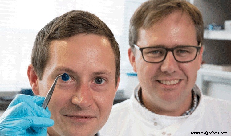
Despite a modest pool of donor corneas, the demand far outstrips supply, prompting researchers to develop artificial corneal substitutes. Newcastle University scientists have now demonstrated a rapid 3‑D printing method that could one day ease this gap.
The cornea—the eye’s outermost, refractive layer—contributes about 80 % of the eye’s focusing power. Engineering a functional substitute therefore demands precise curvature to replicate this optical function.
How It Was Made
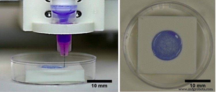
Researchers blended human corneal stromal cells, harvested from healthy donors, with sodium alginate and methacrylated type I collagen to create a “bio‑ink.” This gel is both cell‑friendly and mechanically suitable for extrusion printing.
Using an affordable 3‑D bioprinter, the bio‑ink was deposited in concentric rings to build a cornea‑shaped construct. The entire process completed in just 10 minutes.
Bio‑ink
The collagen‑alginate mix provides enough structural integrity to maintain shape while keeping embedded cells viable at room temperature. The viscosity is tuned so the material can be extruded without compromising cell survival.
With bio‑ink, tissue geometry can be customized to a patient’s eye by scanning the cornea and translating the dimensions into G‑code for the printer. This allows precise replication of complex anatomical features.
The final construct undergoes remodeling in a physiological environment, promoting appropriate structural, functional, and biomechanical maturation.
Reference: ScienceDirect | Newcastle University
Print fidelity was assessed by measuring peripheral and central thickness, while cell viability of encapsulated keratocytes was monitored on day 1 and day 7 post‑printing.
What’s Next?
Although the artificial cornea is not yet ready for clinical use, researchers plan extended in‑vitro and in‑vivo studies to evaluate long‑term safety, stromal cell phenotype, biocompatibility, and support for epithelial cell growth.
Bottom line: it will likely take several years before clinicians can confidently replace a donor cornea with a 3‑D‑printed alternative.
Read: Vision‑Enhancing Nanodrops Could Replace Eyeglasses
Industrial Technology
- How Technology Has Changed Manufacturing of Robots
- Six Sigma: Cut Costs & Boost Customer Satisfaction
- Celebrate Manufacturing Excellence—Choose Your Own Day for Manufacturing Day
- Mastering Tolerance Stacking: A Practical Guide
- Essential Safety Guidelines for Loading and Unloading Silos
- Hyperbolic Functions: Fundamentals and Applications
- How AI Can Reduce Footwear Return Rates and Boost Retail Profitability
- Accelerate Your Production: Proven Tips to Speed Up Manufacturing
- Duo‑Lock™ Modular Systems: Unmatched Solid Carbide Performance
- Enhance PCB Design Efficiency with OrCAD PSpice Simulation