


Magnesium‑Doped Zinc Oxide Nanoparticles: Optimizing Photocatalytic Degradation and Antibacterial Efficacy
Abstract
Using a simple co‑precipitation approach, we synthesized pristine and Mg‑doped ZnO nanoparticles (NPs) with 0–7.5 mol % Mg. X‑ray diffraction confirmed a single hexagonal wurtzite phase, while SEM revealed nanoscale grains (30–110 nm) with hexagonal morphology. Mg incorporation lowered the band gap from 3.36 to 3.04 eV and produced a red‑shifted absorption edge. Photoluminescence showed enhanced visible emission between 430 and 600 nm, indicative of increased defect density. In photocatalytic tests, 7.5 % Mg‑ZnO achieved 78 % degradation of Rhodamine B under UV‑Vis irradiation. Antibacterial assays against Gram‑positive (S. aureus) and Gram‑negative (E. coli, Proteus) strains demonstrated that Mg doping progressively intensified zone‑of‑inhibition diameters, confirming superior antimicrobial performance.
Background
Nanoparticles display size‑dependent properties that enable interactions with biological systems and environmental pollutants. Zinc oxide (ZnO) is a cost‑effective, chemically stable semiconductor widely used in sunscreens, photocatalysis, and antibacterial applications. Doping ZnO with divalent metals such as Mg can tailor its optical band gap, defect chemistry, and surface reactivity, thereby enhancing photocatalytic and antimicrobial activities. Mg, being smaller than Zn, substitutes Zn²⁺ in the lattice, inducing strain and creating oxygen vacancies that serve as active sites for charge‑carrier separation and reactive‑oxygen‑species (ROS) generation.
Methods
Analytical‑grade reagents (Sigma‑Aldrich) were used without further purification. ZnCl₂ and MgCl₂ were mixed in molar ratios of 0.975:0.025, 0.950:0.050, and 0.925:0.075 to obtain 2.5 %, 5 %, and 7.5 % Mg‑doped ZnO, respectively. A 1 M NaOH solution was added dropwise to the metal‑chloride mix under vigorous stirring; the resulting precipitate was aged for 6 h, filtered, washed with deionized water and acetone, dried at 100 °C, ground, and calcined at 300 °C for 4 h. The co‑precipitation route yields high‑purity, nanosized particles at ambient conditions.
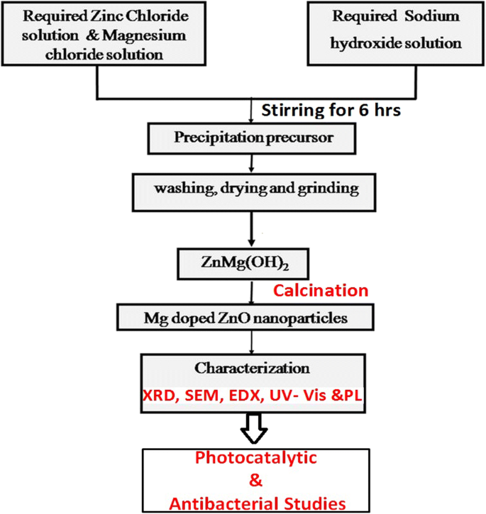
Structural, morphological, and optical properties were characterized by XRD (Bruker D8, Cu Kα, λ = 1.54056 Å), FE‑SEM (ZEISS), UV‑Vis spectroscopy (Hitachi U‑3900H), and photoluminescence (Job HR800, 325 nm He‑Cd laser). Antibacterial activity was assessed via agar disc diffusion against E. coli, S. aureus, and Proteus strains.
Results and Discussion
Structural Analysis
XRD patterns (Fig. 2) exhibited characteristic peaks at 31.8°, 34.5°, 36.3°, 47.5°, 56.7°, 62.9°, and 68°, confirming the hexagonal wurtzite structure (JCPDS 36‑1451). No secondary phases were detected. Peak intensities diminished with increasing Mg, indicating slight crystallinity loss due to lattice distortion. The (101) peak shifted to lower angles, consistent with Mg²⁺ (ionic radius = 0.57 Å) substituting Zn²⁺ (0.60 Å). Crystallite sizes, calculated via Scherrer’s equation, increased from ~35 nm (pure) to ~70 nm (7.5 % Mg). Table 1 summarizes lattice parameters, atomic packing fractions, and strain values.
Morphology and Composition
FE‑SEM images (Fig. 3) show well‑defined hexagonal grains ranging from 30 to 110 nm, with higher Mg content yielding better dispersion. EDS spectra (Fig. 4) confirm the presence of Zn, Mg, and O, with Mg intensity correlating to doping level and no extraneous elements detected.
Optical Properties
UV‑Vis absorption (Fig. 5a) reveals increased absorbance and a red‑shifted absorption edge as Mg content rises. Tauc plots (Fig. 5b) yield band gaps decreasing from 3.36 eV (pure) to 3.04 eV (7.5 % Mg), reflecting enhanced quantum confinement and defect‑induced states.
Photoluminescence
PL spectra (Fig. 6) display a strong UV peak (~400 nm) and a broad visible band (450–620 nm). Visible emission intensifies with Mg doping, indicating more surface anion vacancies and improved electron‑hole separation.
Photocatalytic Performance
Under 125 W (311 nm) UV irradiation, 7.5 % Mg‑ZnO achieved 78 % degradation of Rhodamine B after 120 min, outperforming pure ZnO (≈ 20 %). Kinetic analysis (Fig. 9) shows first‑order rate constants increasing from 1.09 × 10⁻³ to 1.26 × 10⁻² min⁻¹ with Mg content, confirming enhanced charge‑carrier dynamics. The improvement is attributed to increased surface area, oxygen vacancies, and effective electron sinks provided by Mg incorporation.
Antibacterial Activity
Disc diffusion assays (Fig. 10) reveal that Mg‑ZnO NPs inhibit both Gram‑positive and Gram‑negative bacteria. Zones of inhibition enlarge with higher Mg percentages, reaching 18 mm against E. coli and 20 mm against S. aureus for 7.5 % Mg‑ZnO. Enhanced ROS generation and reduced band gap facilitate stronger antimicrobial action.
Conclusions
Mg doping via co‑precipitation produces phase‑pure ZnO NPs with tunable morphology, reduced band gap, and increased defect density. 7.5 % Mg‑ZnO delivers superior photocatalytic degradation of organic dyes and potent antibacterial activity, making it a promising candidate for environmental remediation and antimicrobial applications.
Nanomaterials
- Nanofiber & Filament-Based Nanocarriers: Advancing Precision Drug Delivery
- Nanoparticle-Based Cancer Therapy: Advances, Mechanisms, and Clinical Translation
- Eco‑Friendly Copper Oxide Nanoparticles Doped with Ginger and Garlic Extracts Exhibit Potent Antibacterial Activity Against Escherichia coli
- Enhanced Photocatalytic Performance of ZnO/In₂O₃ Hybrid Nanostructures via Hydrothermal Synthesis
- Ag3PO4/BiFeO3 Heterojunctions: Superior Visible‑Light Photocatalytic Degradation of Acid Orange 7
- Dialysis‑Derived Tadpole and Sphere Hemin Nanoparticles: A 308‑Fold Solubility Boost for Iron Bioavailability
- Optimizing ZnO‑Based Nanohybrids: How Materials, Heterojunctions, and Crystal Orientation Enhance Methyl Orange Degradation
- Partially BiVO4-Modified ZnO Porous Nanosheets: Solar‑Driven Photocatalysis with Superior Charge Separation
- Enhanced Photocatalytic and Antimicrobial Performance of Mg-Doped ZnO Nanorods: Structural Characterization and Molecular Docking Insights
- Quantitative Study of PLGA Nanoparticle Uptake in Laryngeal Cancer and Immune Cells to Improve Drug Delivery