


Magnetic Resonance Imaging (MRI): Principles, Evolution, and Clinical Applications
Magnetic Resonance Imaging (MRI) is a non‑invasive imaging modality that harnesses the natural resonance of hydrogen atoms in a strong magnetic field to produce detailed images of soft and hard tissues. First conceptualized in 1945, MRI has evolved into a cornerstone of modern diagnostics, offering unparalleled contrast resolution without ionising radiation.
History
Early insights into nuclear magnetic resonance (NMR) in the 1900s led to the discovery of atomic magnetic properties. In 1938 I. I. Rabi demonstrated the first NMR apparatus, though it was limited to gaseous samples and indirect measurements. The breakthrough came in 1945 when Felix Bloch and Edward Purcell independently developed robust NMR devices that could probe a variety of systems. By the mid‑1960s, researchers adapted NMR to biological tissues, setting the stage for medical imaging.
The first MRI scanner emerged in 1973 when an NMR‑based imaging technique combined with computer‑generated tomography produced the first diagnostic image of a living mouse. Human studies followed shortly thereafter, and incremental improvements—such as faster gradient switching and higher field strengths—have dramatically shortened scan times and sharpened spatial resolution.
How MRI Works
During an MRI examination the patient is positioned within a powerful, static magnetic field produced by a superconducting or permanent magnet. Radiofrequency (RF) pulses are then applied, tipping the alignment of hydrogen nuclei. When the RF field is switched off, the nuclei relax back to equilibrium, emitting RF signals that are detected by receiver coils. These signals are converted into digital data, processed through Fourier transforms, and reconstructed into two‑ or three‑dimensional images.
Key parameters influencing image contrast include proton density, T1 relaxation time, and T2 relaxation time. By manipulating the timing of RF pulses and gradient fields, radiologists can accentuate differences between tissues, detect pathology, and even assess functional activity.
Clinical Applications
- Neuroimaging – High‑resolution views of brain anatomy and pathology, including tumours, strokes, and demyelinating disease.
- Functional MRI (fMRI) – Real‑time mapping of blood‑oxygen‑level‑dependent signals to study brain activity.
- Musculoskeletal Imaging – Precise assessment of ligaments, cartilage, and bone marrow lesions without ionising radiation.
- Cardiac MRI – Evaluation of cardiac function, perfusion, and tissue characterisation.
– Emerging techniques that detect non‑hydrogen nuclei such as ^13C and ^31P for metabolic imaging.
Core Components
The MRI system comprises several essential elements:
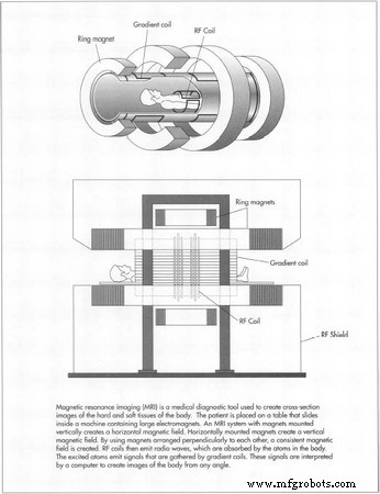
- Magnet – Generates a stable, homogeneous magnetic field. Superconducting magnets (niobium‑titanium coils cooled with liquid helium) dominate clinical scanners due to their low operating cost and high field strengths (1.5 T–3 T). Resistive and permanent magnets are used in research or portable units.
- Gradient Coils – Rapidly alter the magnetic field to encode spatial information. Three orthogonal sets provide three‑dimensional localisation.
- RF System – Transmits excitation pulses and receives emitted signals. Common coil designs include saddle, solenoid, and birdcage arrays, often paired with pre‑amplifiers to maximise signal‑to‑noise ratio.
- Computing & Reconstruction – Dedicated hardware converts analog signals to digital, applies Fourier transforms, and renders images. Advanced software enables parallel imaging and compressed sensing to reduce scan time.
Manufacturing & Assembly
Individual subsystems are fabricated separately and then integrated into a single, often >100 tonne, unit. Key steps include:
- Fabrication of superconducting coils, immersion in liquid helium, and vacuum‑sealed housing.
- Construction of gradient coils with epoxy‑reinforced copper windings and eddy‑current shielding.
- Assembly of RF coils and pre‑amplifiers, ensuring impedance matching and minimal interference.
- Integration of the control computer, user interface, and data storage modules.
- Final on‑site assembly and calibration under strict environmental conditions.
Quality Assurance
Every MRI system undergoes rigorous visual and electrical inspections, performance validation, and environmental stress testing. Manufacturers adhere to guidelines from organisations such as the American College of Radiology (ACR) and the International Electrotechnical Commission (IEC). These protocols confirm field homogeneity, gradient linearity, RF safety, and patient comfort.
The Future of MRI
Ongoing research focuses on:
- Increasing field strengths (e.g., 7 T) to enhance resolution.
- Implementing parallel imaging and compressed sensing to cut scan times.
- Developing real‑time, motion‑robust sequences for dynamic studies.
- Exploring ultrahigh‑field portable scanners for point‑of‑care imaging.
- Integrating AI‑driven reconstruction to further suppress noise and improve diagnostic confidence.
Conclusion
From its origins in fundamental physics to its current status as a versatile diagnostic tool, MRI continues to evolve. Advances in magnet design, signal processing, and clinical protocols promise faster, clearer, and safer imaging for patients worldwide.
Manufacturing process
- Understanding Relay Construction: From Solenoids to Industrial Applications
- Electromagnetism: From Oersted’s Discovery to Modern Applications
- The Evolution of Videotape: Revolutionizing Media & Home Entertainment
- Magnets: Types, Materials, Manufacturing, and Future Applications
- The Evolution of Floppy Disks: From Magnetic Media to Optical Storage
- Heparanase‑Targeted Magnetic Gold Nanoparticle Probe Enhances MRI Detection of Tumor Metastasis
- HER2-Targeted Magnetic Nanosensitizer Enhances In Vivo MRI for HER2-Positive Cancers
- Dual-Targeted Paramagnetic Liposomes for αvβ3 Integrin and NRP‑1: A Powerful MRI Tool for Early Tumor Detection
- Magnetite Nanocluster-Based Theranostic Agents for T2‑Weighted MRI and pH‑Responsive Doxorubicin Delivery
- Understanding Magnetic Chip Conveyors: Function, Features, and Applications