


Lanthanum Strontium Manganite Nanoparticles: Airway Epithelial Cell Toxicity and Mucus Secretion Impact
Abstract
Nanoparticle toxicity research has intensified as these materials become ubiquitous in everyday products. Certain nanoparticles can stimulate mucus secretion, potentially harming airway integrity. Lanthanum strontium manganite (LSM) is a perovskite nanoparticle extensively used in solid‑oxide fuel cells (SOFCs) because of its high electrical conductivity, robust electrochemical activity for the O2 reduction reaction, thermal stability, and long‑term performance. However, limited data exist on LSM’s biological effects. In this study, primary tracheal epithelial cells were exposed to LSM concentrations up to 500 µg mL−1. Results indicated only moderate effects on cell viability, reactive oxygen species (ROS) production, cytochrome C release, and caspase‑3 expression. While apoptosis markers remained largely unchanged, LSM significantly inhibited mucus secretion in a dose‑dependent manner. Thus, although LSM shows low cytotoxicity toward airway cells, its capacity to reduce mucus production could compromise airway clearance.
Introduction
Lanthanum strontium manganite (LSM) possesses a perovskite crystal structure with the general formula La1−xSrxMnO3, where x reflects the strontium doping level. This material can be processed as a powder for tape casting, air/thermal/plasma spray, and fuel‑cell applications. LSM’s high surface area and electrochemical performance make it a leading electrode candidate for SOFCs, which are central to the emerging hydrogen economy. Despite extensive studies on LSM’s mechanical and electrical properties, its biological interactions remain underexplored. Many nanoparticles are known to elevate mucus secretion and contribute to respiratory disorders. Emerging evidence suggests LSM may also serve as a drug carrier or imaging contrast agent, but its potential toxicity must be clarified before any biomedical deployment. This investigation evaluates LSM’s cytotoxic profile in primary tracheal epithelial cells, assessing ROS generation, mitochondrial integrity, apoptosis, and mucus secretion.
Results and Discussions
Nanoparticle Characterization and Cell Viability
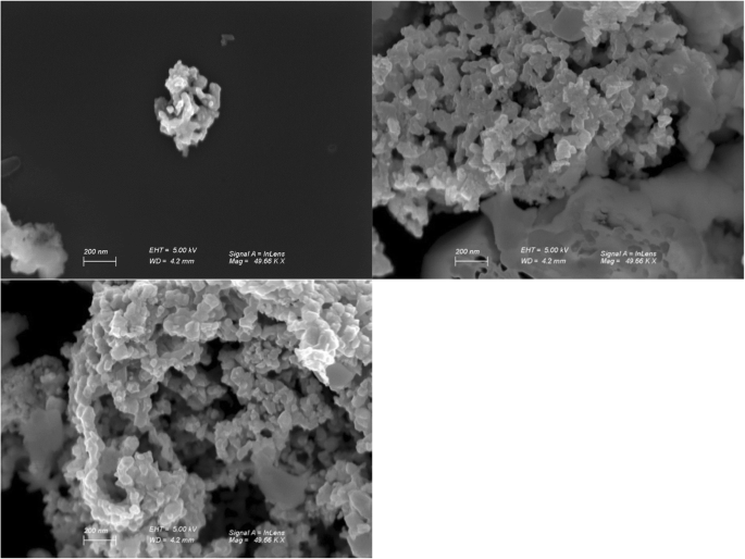
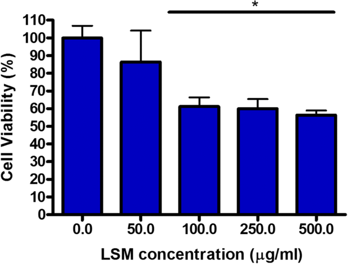
Surface morphology was examined by scanning electron microscopy (SEM). LSM nanoparticles displayed a coarse surface and aggregated into clusters ranging from ~35 nm to >200 nm (Fig. 1). Cell viability was measured with the CCK‑8 assay. Viability sharply decreased between 50 and 100 µg mL−1 but plateaued thereafter, remaining stable at higher concentrations (Fig. 2).
ROS Production and Mucin Release
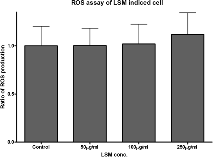
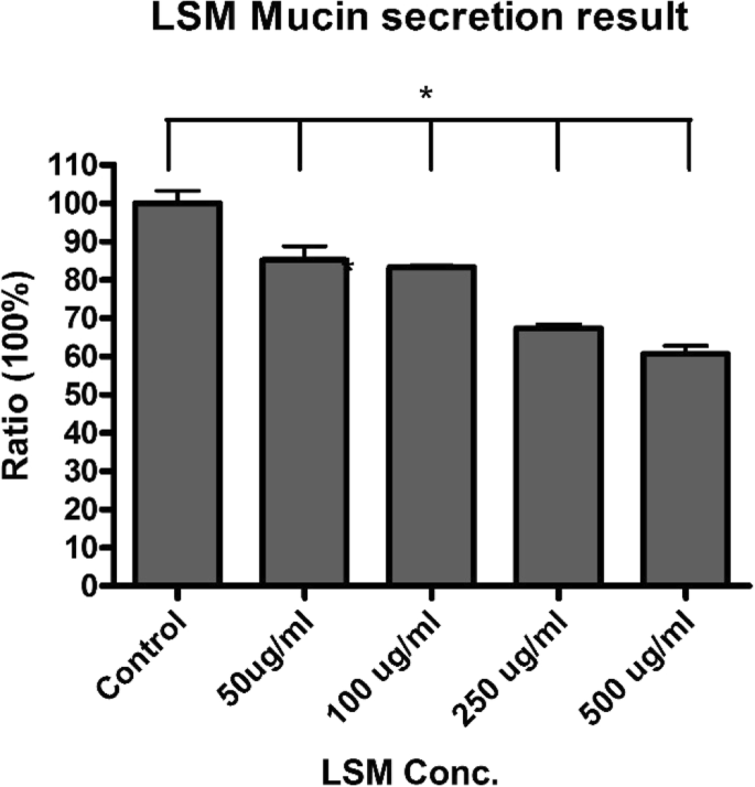
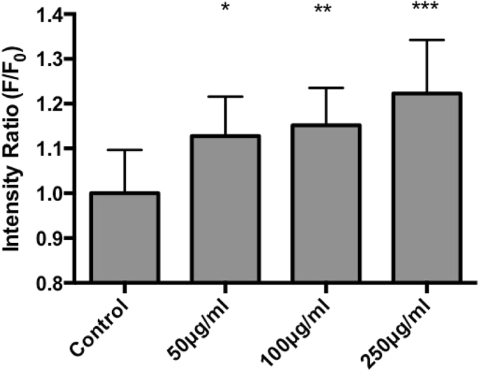
ROS levels were quantified using CM‑H2DCFDA fluorescence. Increasing LSM concentration did not significantly elevate ROS; the 250 µg mL−1 point showed the largest deviation, yet remained statistically insignificant (Fig. 3). Mucin secretion, measured by enzyme‑linked lectin assay (ELLA), decreased progressively with higher LSM doses, dropping to ~40 % of control at 500 µg mL−1 (Fig. 4).
Mitochondrial Integrity and Apoptosis
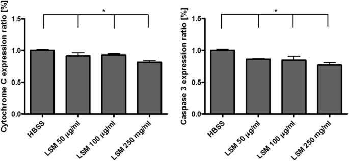
JC‑1 staining revealed mitochondrial depolarization at concentrations ≥100 µg mL−1 (Fig. 5). Despite this, cytochrome C release and caspase‑3 activation only showed modest reductions, indicating limited apoptosis induction (Fig. 6).
Conclusions
This first assessment of LSM toxicity in airway epithelial cells demonstrates negligible cytotoxicity and apoptosis, but a pronounced inhibition of mucus secretion. While ROS generation and mitochondrial damage were minimal, the reduced mucus release may impair airway clearance and warrants careful evaluation for future applications in energy and biomedical fields.
Materials and Methods
Culture of Tracheal Primary Cells
Primary bovine tracheal epithelial cells were isolated per established protocols, cultured in serum‑free medium (SFM) supplemented with EGF 1–53 and bovine pituitary extract (BPE), and maintained on collagen‑coated plates at 37 °C, 5 % CO2. Cell density was monitored via trypan blue exclusion.
Cell Preparation and Viability Assay
Cells were seeded in collagen‑coated 96‑well plates (5 × 10⁴ cells/well) for viability assays and in 4‑well plates (5 × 10⁵ cells/well) for calcium, ROS, and mitochondrial studies. After 24 h incubation, media were replaced with PBS, then with LSM suspensions. Cell viability was quantified with the CCK‑8 assay (WST‑8 conversion to formazan) and expressed as a percentage of untreated control.
Lanthanum Strontium Manganite Nanoparticles
LSM (La0.15Sr0.85MnO3, 35 nm, 99.5 %) was sonicated and tested at 500, 250, 100, and 50 µg mL−1. Concentrations were chosen to match typical TiO2 nanoparticle exposures reported in the literature.
Scanning Electron Microscopy
Particles were drop‑cast on silicon wafers, air‑dried, and imaged with a Zeiss Gemini SEM to confirm size distribution.
Reactive Oxygen Species Assay
ROS production was assessed using CM‑H2DCFDA fluorescence after 15 min LSM exposure. Fluorescence intensity was compared to untreated controls.
Mitochondrial Damage Measurement
JC‑1 dye was used to evaluate mitochondrial membrane potential; red/green fluorescence ratios were measured by fluorescence microscopy at 10‑min intervals.
Mucin Secretion and ELLA
After 15 min LSM exposure, supernatants were collected, centrifuged, and assayed for mucin via ELLA using WGA‑HRP and TMB substrate. Absorbance was read at 450 nm.
Apoptosis Marker Assays
Cell lysates were prepared with RIPA buffer and probed for cytochrome C and active caspase‑3 using ELISA protocols. Secondary HRP‑conjugated antibodies facilitated detection.
Statistical Analysis
Data are presented as mean ± SD from at least three independent experiments. One‑way ANOVA determined significance (p < 0.05).
Abbreviations
- BPE
- Bovine pituitary extract
- ELISA
- Enzyme‑linked immunosorbent assay
- LSM
- Lanthanum strontium manganite
- ROS
- Reactive oxygen species
- SFM
- Serum‑free medium
- SOFCs
- Solar oxidized fuel cells
Nanomaterials
- High‑Efficiency, Low‑Cost Perovskite Solar Cells: Progress, Challenges, and Future Directions
- Effects of 15‑nm Gold Nanoparticles on Proliferation, Apoptosis, and Spheroid Formation in HT29 Colon Carcinoma and SPEV Embryonic Kidney Cells
- Using GaN/Fe Nanoparticles to Magnetically Guide Endothelial Cells in Vitro
- High‑Efficiency Broadband Solar Absorber Using Tungsten Nanoparticle Multilayers
- PEG-CoFe₂O₄ Nanoparticles: Assessing Toxicity and Curcumin’s Protective Effect
- Optimizing Perovskite Solar Cell Efficiency with Size‑Controlled Ag Nanoparticles in a TiO₂ Compact Layer
- Targeted Lipid Nanoparticles Functionalized with Transferrin Enhance Paclitaxel Efficacy in Leukemia Cells
- Efficient Ambient‑Air Fabrication of Mesoporous Perovskite Solar Cells Using N‑Butyl‑Amine‑Enhanced PbI₂ Precursors
- Comparative Toxicity of PEG-Coated Cobalt Ferrite Nanoparticles and Nanospheres
- Enhanced Stability and Efficiency of 2D Perovskite Solar Cells Through Bromine Incorporation